Can standardized CT scans improve safety?

Dr. Diana Miglioretti and University of California colleagues are developing and testing a new way to lower radiation doses while still producing sharp images
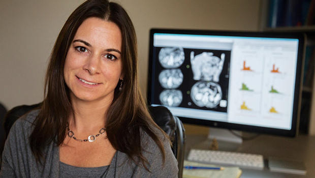
By Diana L. Miglioretti, PhD, a senior Investigator at Kaiser Permanente Washington Health Research Institute and the Dean’s professor of biostatistics at the University of California, Davis School of Medicine Department of Public Health Sciences.
Use of computed tomography (CT) scans keeps rising, and when you get a CT scan, you’re exposed to much more ionizing radiation than if you got an X-ray. It’s a balancing act: The more radiation, the clearer the image — but also the more potential damage to DNA, which can actually cause cancer, especially in people who are more susceptible.
This is a paradox: CT is an excellent way to detect diseases including cancer early, quickly, and accurately. But some studies, including ours, have predicted that repeated high-dose CT scans may make future cancers more common.
I want patients to feel confident that they’re getting the lowest radiation doses needed for effective imaging. For that to happen, we need consistent standards — and less variation in dose for no particular reason.
Work started at KPWA
That’s why I first started working to standardize this balancing act in medical imaging at Kaiser Permanente Washington. We systematically shared best practices. And we gave radiology professionals feedback that let them compare patients’ radiation exposure from imaging exams they did with exams their peers did.
Now my colleagues at the University of California (UC) and I have used this approach to help standardize radiation in CT scans at the five medical centers that make up UC Health: UC Davis (where I work), UC Irvine, UC Los Angeles, UC San Diego, and UC San Francisco.
With coauthors including my longtime colleague Rebecca Smith-Bindman, MD, of UCSF, we recently published our results in JAMA Internal Medicine: Optimizing Radiation Doses for Computed Tomography Across Institutions.
After studying 160,000 CT scans at these medical centers over more than a year, we decided to focus on CT scans of the chest, abdomen, and head. These scans account for more than 80 percent of all CT imaging. We found that after our simple intervention, the mean effective radiation dose for abdominal CT scans was lowered by 25 percent; and for standard chest CT scans, by 19 percent. Doses for head CT scans varied less after the intervention, meaning both high and low doses were brought closer to the mean. (Low doses can lead to “noisy” images that are hard to interpret, which could require re-imaging and thus doubling the radiation exposure.)
Projecting from these results, we estimate that applying this approach to all abdominal CT scans in the United States could result in 12,000 fewer cancers a year. And that doesn’t count chest, head, and other scans.
We are now conducting a much larger trial including over 125 facilities from the United States and other countries, which will tell us even more. And ultimately, we hope the work we began years ago in Washington will result in fewer cancers and many lives saved.
Learn more about Kaiser Permanente Washington Health Research Institute. Sign up for our free monthly newsletter.


